
One of the first things we tried (with Yujen and Lily) was the protocol from Rockwood et al. to make micro- and nano-scale spheres via phase separation of silk fibroin suspended in polyvinyl alcohol (PVA). The synthesis steps were relatively simple – mix SF and PVA solutions in recommended ratios, pour them out in a wide-bottom beaker and let them dry overnight into a film, rehydrate the film and put it on a shaker to dissolve the PVA and liberate the silk micro/nano-spheres, centrifuge to pellet just the spheres, and finally resuspend in water.
With some help from Aaron Straight over in Biochemistry we made it through the process and I prepared some samples for scanning electron microscopy (SEM) by simply depositing some of the final sphere suspension on silicon wafer chips:
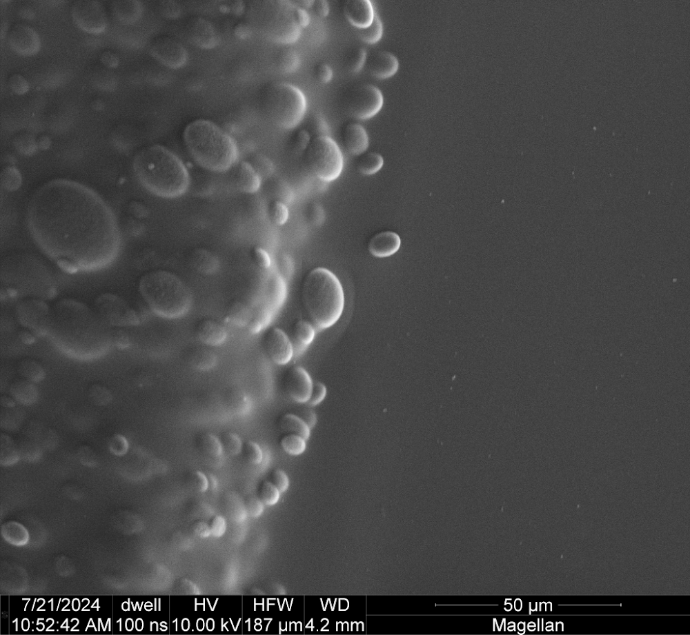
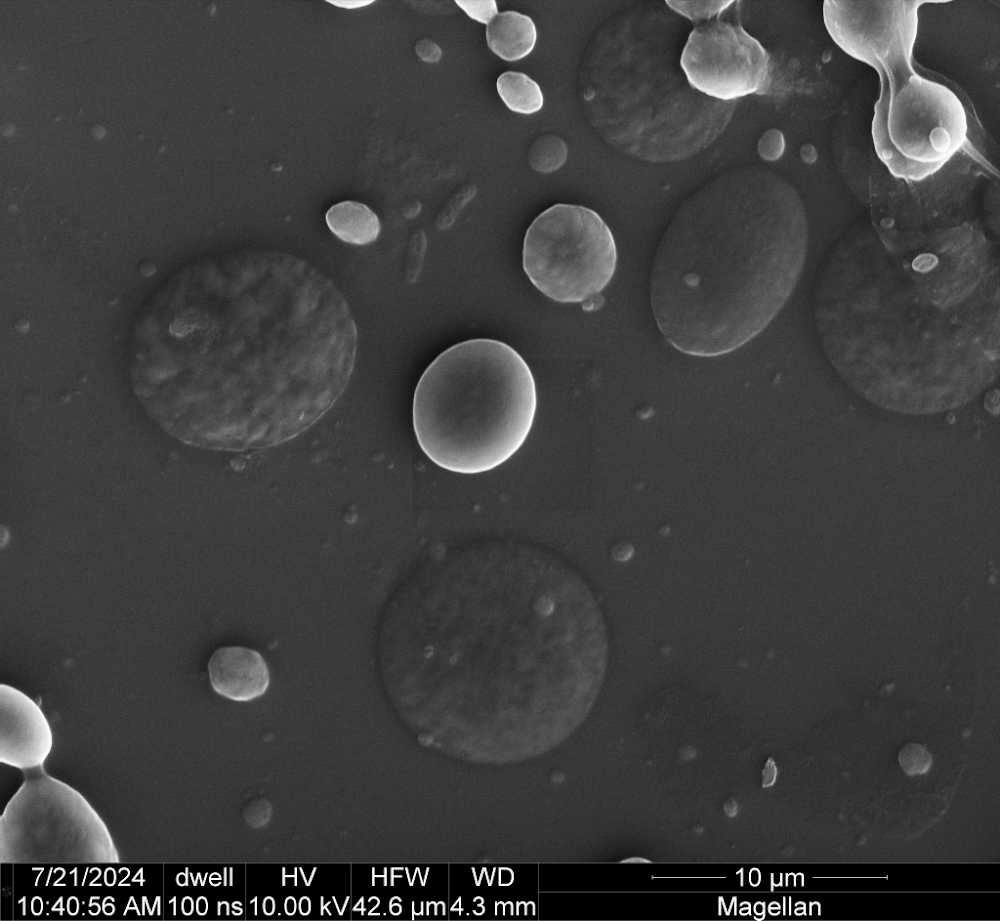
I think the image on the left shows a clump of silk spheres still stuck in a flake of undissolved PVA. The image on the right shows some isolated silk spheres (the bright egg-looking structures, such as in the near center of the frame) together with what I think must be some spheres that collapsed into silk pancakes (the dark/gray lumpy round blobs, such as just below and to the left of the bright egg). In the image on the right you can see what appear to be a very broad range of sizes of silk spheres, with the central bright egg looking like ~5-7 microns in diameter and some of the things that appear just as round specks (assuming they are in fact smaller spheres) well below 1 micron in diameter.
We haven’t really used these micro/nano-spheres for anything but this was a fun first exercise in the material affordances of liquified silk fibroin. Early on I had some thoughts about trying to use silk casting to make really small structures that would be shown via microscope photography, but I have since moved away from that…
